
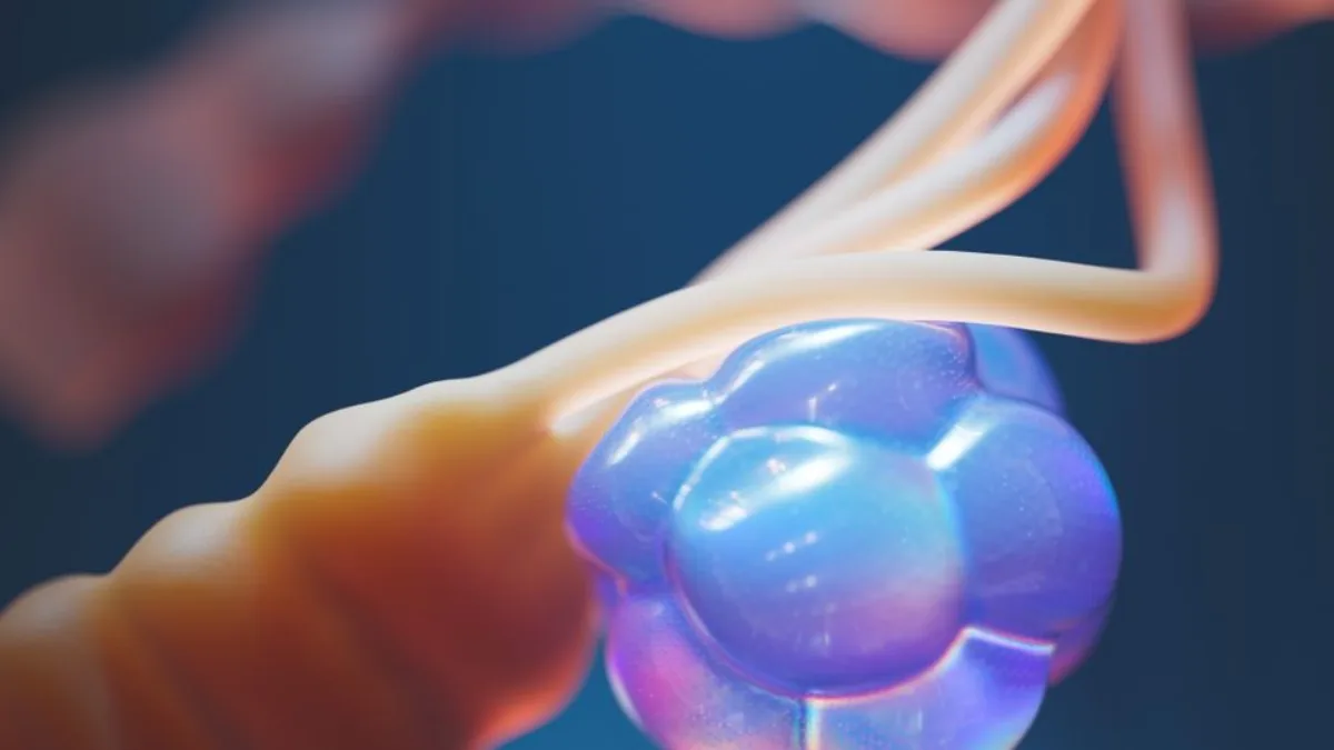
The intricate architecture of chromosomes, far beyond the simple double helix, underpins the fundamental processes of life. Understanding their structural hierarchy, spatial organization, and dynamic epigenetic modifications is crucial for deciphering gene regulation, cellular identity, and disease pathogenesis. This exploration delves into the advanced structural biology, spatial organization, and epigenetic mechanics of chromosomes, moving beyond textbook definitions to reveal their complex functional interplay.
Quick Summary / Key Takeaway
Chromosomes are hierarchically packaged DNA structures. Their function is dictated by histone modifications, specialized domains like TADs, and nuclear compartmentalization. Advanced techniques map these structures, revealing insights into non-canonical inheritance and genomic instability.
Structural Entity & Macromolecular Architecture
The journey from a linear DNA double helix to a compact metaphase chromosome involves a remarkable series of hierarchical packaging events. This process is orchestrated by a complex interplay of proteins and DNA sequences, ensuring the faithful transmission of genetic information while allowing for dynamic regulation of gene expression. At the most fundamental level, the DNA double helix is wrapped around a core of histone proteins.
The Histone Core Octamer and PTMs
The fundamental unit of chromatin packaging is the nucleosome, formed by approximately 147 base pairs of DNA wrapped around a histone octamer core. This core is composed of two copies each of the four core histone proteins: H2A, H2B, H3, and H4. This fundamental interaction between the negatively charged DNA and the positively charged histone proteins forms the basis of chromatin condensation.
The N-terminal tails of these histones, which protrude from the nucleosome core, are extensively modified by a diverse array of post-translational modifications (PTMs). These PTMs, including acetylation, methylation, phosphorylation, and ubiquitination, act as a critical regulatory layer.
They influence the accessibility of DNA to transcription factors and other regulatory proteins, thereby dictating the local chromatin state. For instance, histone acetylation, particularly on H3 and H4 tails, generally correlates with transcriptional activation by neutralizing the positive charge of histones, loosening their interaction with DNA.
Conversely, specific methylation marks, such as H3K9me3, are strongly associated with gene silencing and the formation of heterochromatin. These modifications are not static; they are dynamically added and removed by specific enzymes, creating a complex “histone code” that is interpreted by the cellular machinery.
Centromeres and CENP-A Kinetochore Recruitment
Centromeres are specialized chromosomal regions essential for accurate chromosome segregation during cell division. They serve as the assembly platform for the kinetochore, a proteinaceous structure that mediates the attachment of chromosomes to spindle microtubules.
While centromeric DNA sequences can vary significantly between species, their epigenetic identity is conserved. This epigenetic definition is primarily driven by the presence of a unique histone H3 variant, CENP-A (Centromere Protein A). CENP-A replaces canonical H3 in nucleosomes specifically at centromeres. This substitution is crucial for recruiting other kinetochore proteins, forming a functional kinetochore complex.
The deposition and maintenance of CENP-A are tightly regulated, ensuring that only the correct centromeric regions are epigenetically marked. The kinetochore machinery, assembled on the CENP-A nucleosomes, is a multi-protein complex that spans the centromeric chromatin and interacts with microtubules.
This interaction ensures that each daughter cell receives a complete set of chromosomes during mitosis and meiosis. Defects in CENP-A deposition or kinetochore assembly can lead to aneuploidy, a hallmark of many cancers and developmental disorders.
Telomeres and the Shelterin Complex
Telomeres are repetitive DNA sequences located at the ends of linear chromosomes. Their primary function is to protect the chromosome ends from being recognized as DNA damage sites by cellular repair machinery. Without telomeres, the cell would perceive chromosome ends as double-strand breaks, triggering inappropriate repair pathways that could lead to chromosome fusions and genomic instability.
The repetitive sequence of telomeres, typically TTAGGG in vertebrates, is maintained by the enzyme telomerase. However, the physical protection of these ends is largely conferred by a specialized protein complex known as Shelterin. Shelterin is a six-protein complex that binds to telomeric DNA, forming a protective cap.
The core components of Shelterin include TRF1 and TRF2, which directly bind to telomeric repeat sequences. These are further stabilized by accessory proteins like RAP1, TIN2, TPP1, and POT1. This complex effectively shields the single-stranded 3′ overhang of the telomere, preventing its recognition by DNA repair proteins such as ATM and ATR. This shielding mechanism is critical for maintaining genomic integrity and preventing cellular senescence or apoptosis.
High-Order Functional Domains and Spatial Organization
Beyond the linear arrangement of DNA and its packaging into nucleosomes, chromosomes exist within a highly organized three-dimensional space inside the nucleus. This spatial organization is not random; it reflects functional compartmentalization and plays a critical role in regulating gene expression and genome stability. Understanding this nuclear architecture requires examining how chromatin is organized into higher-order structures.
Topologically Associating Domains (TADs) and Loop Extrusion
Topologically Associating Domains (TADs) represent fundamental structural and functional units of the genome. These are genomic regions, typically ranging from 100 kilobases to 1 megabase in size, that exhibit significantly higher frequencies of intra-domain DNA-DNA interactions compared to inter-domain interactions.
TADs act as regulatory neighborhoods, insulating genes within them from the regulatory elements of neighboring TADs. The formation and maintenance of TADs are largely mediated by the process of loop extrusion. This mechanism involves the cohesin complex, a ring-like protein structure that encircles DNA.
Cohesin, often guided by the CCCTC-binding factor (CTCF), can extrude DNA loops, effectively tethering distant genomic regions together. CTCF binding sites, often found at the boundaries of TADs, play a crucial role in defining these domains.
When cohesin encounters opposing CTCF-bound orientations at TAD boundaries, loop extrusion is halted, thereby establishing the TAD structure. This organization ensures that enhancers within a TAD preferentially interact with their cognate promoters, while preventing inappropriate cross-talk with other regulatory elements.
A/B Compartments and Phase Separation
At a larger scale, the interphase nucleus is organized into distinct compartments, broadly categorized as A and B compartments. These compartments are identified through genome-wide contact frequency analyses, such as Hi-C. A-compartments are generally characterized by being gene-dense, transcriptionally active, and enriched in euchromatin.
They tend to localize to the interior of the nucleus. In contrast, B-compartments are typically gene-poor, transcriptionally repressed, and enriched in heterochromatin. These regions are often associated with the nuclear lamina, forming Lamina-Associated Domains (LADs). The segregation into A and B compartments reflects a broad organizational principle that segregates active and inactive genomic regions.
The formation of these compartments and other nuclear bodies is increasingly understood through the lens of liquid-liquid phase separation (LLPS). Chromatin, along with associated proteins, can demix from the nucleoplasm, forming distinct, membrane-less compartments.
This phase separation is driven by multivalent interactions between intrinsically disordered regions of proteins and specific chromatin modifications. It allows for the efficient concentration of regulatory factors and the compartmentalization of distinct nuclear functions.
Comparative Taxonomy of Chromatin States
Chromatin can be broadly classified into distinct states based on its transcriptional activity and epigenetic marks. Understanding these states is fundamental to comprehending chromosome function. Euchromatin is the less condensed form of chromatin, characterized by open DNA accessibility and active gene transcription.
It is typically enriched in histone modifications associated with gene activation, such as H3K4me3 and histone acetylation. Constitutive heterochromatin, on the other hand, is highly condensed and transcriptionally silent throughout the cell cycle.
It is characterized by specific repressive histone marks, most notably H3K9me3, which are recognized by heterochromatin protein 1 (HP1) and other silencing factors. This form of chromatin is crucial for maintaining the stability of repetitive elements like centromeres and telomeres.
Facultative heterochromatin represents a dynamic state that can switch between an open and closed conformation. It is found in regions that are transcriptionally repressed in specific cell types or developmental stages but can be activated under different conditions.
This state is often marked by H3K27me3, a modification established by the Polycomb Repressive Complex 2 (PRC2). The interplay between these chromatin states dictates the accessibility of the genome and the regulation of gene expression.
Advanced Clinical and Functional Mechanics
The complexity of chromosome structure and organization extends to non-canonical forms and intricate regulatory trade-offs that have profound implications for health and disease. Understanding these advanced mechanics provides critical insights into phenomena such as cancer evolution and the origins of genetic variation.
Non-Canonical and Extrachromosomal Inheritance
While the vast majority of genetic material resides within the nucleus in the form of linear chromosomes, several non-canonical forms of genetic material exist and play significant roles. Extrachromosomal DNA (ecDNA) refers to circular or linear DNA molecules that exist outside of the host chromosomes.
These can arise from various mechanisms, including gene amplification events. In cancer, ecDNA is increasingly recognized as a major driver of intratumoral heterogeneity and rapid drug resistance. The ability of ecDNA to amplify genes that confer survival advantages, such as oncogenes or drug targets, allows cancer cells to rapidly adapt to selective pressures.
The extrachromosomal nature of these elements facilitates their independent replication and segregation, leading to high copy numbers and rapid acquisition of new mutations. Beyond ecDNA, other non-canonical chromosomal structures include B-chromosomes and holocentric chromosomes.
B-chromosomes are extra chromosomes found in some species that are not essential for survival but can influence phenotype. Holocentric chromosomes, unlike monocentric chromosomes with a single centromere, have their centromeric activity distributed along their entire length, which impacts their segregation dynamics. These variations highlight the diverse evolutionary strategies for organizing and transmitting genetic information.
Direct Regulatory Trade-offs in Transcription and Replication
The dynamic processes of transcription and DNA replication, both essential for cellular function, can sometimes interfere with each other, leading to significant regulatory trade-offs. One critical area of conflict arises from head-on transcription-replication collisions.
When a replication fork moves in the opposite direction to a transcription complex, it can lead to the formation of R-loops. R-loops are three-stranded nucleic acid structures where a DNA:RNA hybrid is formed, with a displaced single strand of DNA.
These structures can stall replication forks, increase the likelihood of DNA double-strand breaks, and trigger genomic instability. The cell has evolved intricate mechanisms to resolve R-loops, involving specific RNA helicases and nucleases.
However, the constant potential for these collisions represents a fundamental challenge in genome maintenance. Furthermore, the spatial organization of chromatin itself creates inherent trade-offs. For example, highly condensed heterochromatin, while essential for stability, renders the DNA inaccessible to transcription machinery, effectively silencing genes.
Conversely, the open nature of euchromatin facilitates gene expression but may also make the DNA more vulnerable to damage. The cell must constantly balance these competing demands, employing dynamic epigenetic mechanisms to regulate chromatin accessibility and ensure both faithful replication and appropriate gene expression.
Methodological & Analytical Frameworks
The advanced understanding of chromosome structure and function described above would be impossible without sophisticated high-throughput molecular tools. These technologies allow researchers to probe chromosome conformation, chromatin accessibility, and protein-DNA interactions at a genome-wide scale, providing unprecedented resolution into the dynamic organization of the genome.
Chromosome Conformation Capture: Hi-C and Micro-C
Chromosome Conformation Capture (3C) and its high-throughput derivatives, such as Hi-C and Micro-C, are powerful techniques for mapping the three-dimensional organization of the genome within the nucleus.
The fundamental principle involves cross-linking DNA-protein interactions in situ, followed by restriction enzyme digestion, ligation of proximal DNA fragments, and high-throughput sequencing. Hi-C, in particular, utilizes formaldehyde cross-linking to capture physical proximity between DNA segments.
After cross-linking, the DNA is digested, and the resulting fragments are ligated in dilute conditions, favoring intra-molecular ligation of physically interacting DNA segments. These ligated junctions are then sequenced to generate a contact map, illustrating the frequency of interactions between different genomic loci.
Micro-C is a refinement of Hi-C that uses micrococcal nuclease (MNase) for DNA digestion, allowing for the capture of chromatin structure at a finer resolution, down to nucleosome-level organization.
These methods have been instrumental in identifying TADs, A/B compartments, and other large-scale organizational features of the genome, revealing how linear DNA sequences are folded into complex three-dimensional structures.
Mapping Chromatin Accessibility and Binding: ATAC-seq and ChIP-seq
To understand the functional implications of chromatin organization, it is essential to map regions of open chromatin and the binding sites of specific proteins. Assay for Transposase-Accessible Chromatin using sequencing (ATAC-seq) is a widely used method for identifying accessible regions of the genome.
It leverages the Tn5 transposase, an enzyme that preferentially inserts sequencing adapters into open, nucleosome-depleted DNA regions. This method is highly efficient and requires relatively few cells, making it suitable for various applications.
The resulting sequencing data reveals regions of the genome that are actively being transcribed or are poised for transcription, as these are generally found in a more open chromatin state. Chromatin Immunoprecipitation sequencing (ChIP-seq) is another cornerstone technique used to map the location of specific proteins, such as transcription factors or histone modifications, across the genome.
In ChIP-seq, cells are treated with formaldehyde to cross-link proteins to DNA. The chromatin is then fragmented, and an antibody specific to the protein of interest is used to immunoprecipitate the protein-DNA complexes. After reversing the cross-links, the DNA fragments are sequenced.
The resulting data reveals the genomic regions where the target protein or modification is enriched, providing critical insights into gene regulation, epigenetic states, and the functional landscape of the chromosome. By combining data from ATAC-seq and ChIP-seq, researchers can build a comprehensive picture of how chromatin accessibility and protein binding correlate with transcriptional activity and nuclear organization.
Frequently Asked Questions
What are the basic building blocks of chromosomes?
Chromosomes are built from DNA wrapped around histone proteins, forming nucleosomes. These nucleosomes are further condensed into higher-order structures, ultimately forming the compact chromosome visible during cell division.
How does histone modification affect chromosome function?
Histone post-translational modifications (PTMs) like acetylation and methylation alter chromatin structure, influencing DNA accessibility. This dictates whether genes are actively transcribed or silenced, thereby controlling cellular function.
What is the role of CENP-A in centromeres?
CENP-A is a specialized histone H3 variant that epigenetically defines centromeres. It serves as the foundation for recruiting the kinetochore machinery, essential for accurate chromosome segregation during cell division.
How do TADs contribute to genome organization?
Topologically Associating Domains (TADs) are structural blocks that insulate genomic regions. They promote specific enhancer-promoter interactions within the domain while preventing cross-talk with neighboring regulatory elements.
What is the significance of ecDNA in cancer?
Extrachromosomal DNA (ecDNA) in cancer cells can rapidly amplify oncogenes or drug resistance genes. This extrachromosomal nature allows for quick adaptation and evolution, driving tumor progression and treatment failure.
Which techniques map 3D chromosome interactions?
Techniques like Hi-C and Micro-C are used to map 3D chromosome interactions. They capture physical proximity between DNA segments within the nucleus, revealing genome folding patterns.