
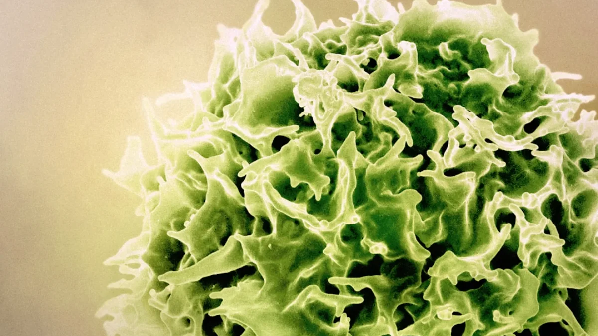
Understanding the fundamental differences between plant and animal cells is essential for grasping how life diverges at the microscopic level. While both are eukaryotic, their structural variations dictate how organisms grow, eat, and interact with their environments. This guide provides a deep dive into the specialized organelles and mechanical systems that define these two distinct biological lineages.
Quick Summary / Key Takeaway
Plant cells possess rigid cellulose cell walls, large central vacuoles, and chloroplasts for autotrophic energy production. Animal cells lack these structures, utilizing flexible plasma membranes and centrioles instead. While both share mitochondria for respiration, plant cells prioritize structural stability, whereas animal cells prioritize mobility and rapid response.
Fundamental Structural Differences: The Outer Boundary
The most immediate distinction between plant and animal cells lies in their external architecture. This boundary determines not only the shape of the cell but also how it handles physical stress and environmental changes. Plant cells are encased in a sturdy exterior, while animal cells maintain a more fluid and adaptable perimeter.
The Plant Cell Wall: Composition and Function
Plant cells are defined by a rigid cell wall located outside the plasma membrane. This structure is primarily composed of cellulose, a complex carbohydrate that provides immense tensile strength. Hemicellulose and pectin act as a matrix, binding the cellulose fibers together to create a tough, protective layer. The cell wall serves as a skeletal framework for the entire plant. It allows plants to grow to great heights without a bony skeleton by supporting the weight of the organism. Furthermore, the wall prevents osmotic lysis, ensuring the cell does not burst when water enters via osmosis.
The Animal Cell Membrane: Flexibility and Glycocalyx
Animal cells lack a cell wall, leaving the plasma membrane as their primary outer boundary. This membrane is a fluid mosaic of phospholipids, proteins, and cholesterol. The absence of a rigid wall allows animal cells to adopt various shapes, which is critical for movement and specialized tissue formation. Many animal cells are covered by a glycocalyx, a carbohydrate-rich coating. This layer facilitates cell-to-cell recognition and provides a level of protection against mechanical damage. Because they lack a wall, animal cells are more susceptible to osmotic changes and must live in nearly isotonic environments.
Energy Production: Photosynthesis vs. Respiration
The way these cells acquire and process energy represents a major evolutionary divergence. While both lineages require ATP to power cellular functions, the source of that energy and the organelles involved differ significantly. This section clarifies the roles of chloroplasts and mitochondria.
Chloroplasts: The Powerhouses of Photosynthesis
Plant cells are autotrophic, meaning they produce their own food using sunlight. This process occurs within chloroplasts, specialized organelles containing the green pigment chlorophyll. Chloroplasts capture light energy to convert carbon dioxide and water into glucose, a form of chemical energy. Chloroplasts have a complex internal structure consisting of thylakoids stacked into grana. This arrangement maximizes the surface area for light absorption. Animal cells lack chloroplasts entirely and must obtain energy by consuming other organisms, a lifestyle known as heterotrophy.
Mitochondria: The Universal Energy Converters
Mitochondria are often called the “powerhouses of the cell” because they perform cellular respiration. In this process, glucose is broken down in the presence of oxygen to produce ATP. Both plant and animal cells contain mitochondria, as both require a way to release energy from stored sugars. In animal cells, mitochondria are the primary source of ATP. They are highly abundant in energy-demanding tissues like muscle. While plants produce glucose via photosynthesis, they still rely on mitochondria to convert that glucose into usable ATP for growth and maintenance.
The Role of Mitochondria in Plant Cells
A common misconception is that plants only use chloroplasts while animals only use mitochondria. In reality, plant cells use both. During the day, chloroplasts produce sugar, but at night or in non-photosynthetic tissues like roots, the cell must rely on mitochondria for energy. Mitochondria in plants are essential for metabolic pathways that occur outside of photosynthesis. They provide the energy needed for nutrient uptake, protein synthesis, and cellular repair. This dual-organelle system allows plants to be highly efficient energy managers in varying environmental conditions.
The Vacuolar System: Storage, Support, and Degradation
Vacuoles are membrane-bound sacs used for storage and transport, but their size and function differ drastically between the two cell types. In plants, the vacuole is a dominant structural feature, whereas in animals, vacuoles are smaller and more specialized.
The Plant Central Vacuole: A Multifunctional Organelle
A mature plant cell typically contains one large central vacuole that can occupy up to 90% of the cell’s volume. This vacuole is surrounded by a specialized membrane called the tonoplast. It stores water, ions, nutrients, and waste products, acting as a reservoir for the cell. The most critical function of the central vacuole is maintaining turgor pressure. By pumping solutes into the vacuole, the cell draws in water via osmosis. This internal pressure pushes against the cell wall, keeping the plant upright and preventing wilting.
Animal Vacuoles and Lysosomes: Specialized Roles
Animal cells contain multiple small, often transient vacuoles or vesicles. These are primarily used for transporting materials into or out of the cell (endocytosis and exocytosis). Unlike the plant vacuole, they do not provide structural support or maintain pressure. In animal cells, the role of waste degradation is handled by lysosomes. These organelles contain digestive enzymes that break down macromolecules and worn-out cell parts. While plants have vacuolar enzymes that perform similar tasks, the animal lysosome is a more specialized compartment for intracellular digestion.
Intercellular Communication: Channels and Connections
Multicellular organisms require efficient communication between cells to coordinate growth and physiological responses. Because plant cells are separated by thick walls and animal cells are not, they have evolved entirely different mechanisms for sharing information and materials.
Plasmodesmata: Direct Cytoplasmic Bridges
Plant cells communicate through plasmodesmata, which are microscopic channels that penetrate the cell walls. These channels are lined by the plasma membrane, creating a continuous cytoplasmic bridge between adjacent cells. This network is known as the symplast. Plasmodesmata allow for the passage of water, ions, and even larger molecules like proteins and RNA. A specialized structure called the desmotubule, derived from the endoplasmic reticulum, often runs through the center of the channel, facilitating the transport of lipids and signaling molecules.
Gap Junctions: Proteinaceous Pores
Animal cells utilize gap junctions for rapid communication. These are clusters of transmembrane proteins called connexons that align between two adjacent cells to form a pore. Unlike plasmodesmata, gap junctions do not involve the sharing of a continuous plasma membrane. Gap junctions allow for the passage of small molecules and ions, which is vital for electrical signaling in heart and nerve tissues. Because animal cells lack a cell wall, these junctions can form and dissolve relatively quickly, allowing for more dynamic tissue remodeling compared to plants.
Cytokinesis and Cell Division
The process of cell division, specifically cytokinesis, is heavily influenced by the presence or absence of a cell wall. While the early stages of mitosis are similar in both cell types, the physical separation of the daughter cells requires different mechanical solutions.
Plant Cytokinesis: The Cell Plate
In plant cells, the rigid cell wall prevents the cell from simply pinching in half. Instead, during late anaphase and telophase, vesicles from the Golgi apparatus congregate at the center of the cell. These vesicles fuse to form a structure called the cell plate. The cell plate grows centrifugally—from the center outward—until it reaches the existing cell walls. Once the plate is complete, it matures into a new primary cell wall, effectively partitioning the original cell into two. This process ensures that both daughter cells maintain structural integrity.
Animal Cytokinesis: The Cleavage Furrow
Animal cells divide through a process called cleavage. A contractile ring of actin and myosin filaments forms just beneath the plasma membrane at the cell’s equator. As these filaments contract, they pull the membrane inward, creating a visible indentation known as the cleavage furrow. The furrow deepens centripetally—from the outside inward—until the parent cell is eventually pinched into two separate daughter cells. This method is highly efficient for cells that lack a rigid exterior and allows for the rapid proliferation seen in animal development.
Advanced Nuances and Biological Edge Cases
While the general rules for plant and animal cells hold true for most species, biology is full of exceptions. Exploring these edge cases provides a more sophisticated understanding of cellular evolution and the diversity of eukaryotic life.
Centrioles: An Exception in Lower Plants
In most textbooks, centrioles are listed as exclusive to animal cells, where they help organize the spindle fibers during division. However, “lower” plants such as mosses, liverworts, and ferns actually possess centrioles and flagellated sperm cells. As plants evolved to live in drier environments, they largely lost the need for flagella and the centrioles that anchor them. Most gymnosperms and all angiosperms (flowering plants) lack centrioles entirely, using alternative microtubule-organizing centers to manage cell division.
Non-Photosynthetic Plant Cells
Not all plant cells are green or capable of photosynthesis. Root cells, for example, live underground and never see sunlight. These cells lack functional chloroplasts but contain other types of plastids, such as amyloplasts, which are specialized for storing starch. Similarly, the cells in flower petals may contain chromoplasts, which store pigments other than chlorophyll to attract pollinators. This demonstrates that while the *potential* for chloroplasts is a defining trait of plants, individual cells adapt their organelle complement based on their specific function.
Functional Equivalency of Lysosomes
While animal cells have distinct lysosomes, plant cells utilize their central vacuole to perform many of the same tasks. The vacuole contains various hydrolytic enzymes that break down proteins, nucleic acids, and lipids, effectively acting as a giant lysosome. In some specialized plant tissues, smaller vacuoles known as “lytic vacuoles” exist specifically for degradation. This highlights a key theme in cell biology: different structures can evolve to perform the same essential function, depending on the overall architecture of the cell.
Frequently Asked Questions
Do plant cells have mitochondria?
Yes, plant cells contain mitochondria to perform cellular respiration. While chloroplasts produce glucose, mitochondria are necessary to convert that glucose into ATP, especially at night or in non-green tissues like roots.
Why do animal cells lack a cell wall?
Animal cells lack a cell wall to allow for mobility and flexibility. This absence enables cells to crawl, change shape, and form complex tissues like muscles and nerves that require movement and contraction.
What is the role of the central vacuole?
The central vacuole stores water, nutrients, and waste. Its primary structural role is maintaining turgor pressure, which pushes against the cell wall to keep the plant rigid and upright.
How do plant cells communicate?
Plant cells communicate through plasmodesmata, which are membrane-lined channels that cross the cell walls. These channels allow for the direct exchange of cytoplasm, ions, and signaling molecules between adjacent cells.
Do all plant cells have chloroplasts?
No, only cells in the green parts of a plant, like leaves and stems, typically contain chloroplasts.